18F-PSMA-1007 PET/CT Improves Staging Vs MRI in Prostate Cancer
Findings support the use of 18F-PSMA-1007 PET/CT in the preoperative workflow of intermediate- and high-risk prostate cancer.


“These findings support the use of 18F-PSMA-1007 PET/CT in the preoperative workflow of intermediate-and high-risk tumors,” according to Nikhile Mookerji, MD.
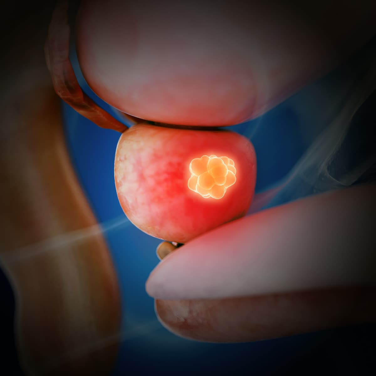
The use of 18F-PSMA-1007 PET/CT showed improved results for the locoregional staging of prostate cancer compared with multiparametric MRI, according to a presentation from the 2024 American Urological Association’s Annual Meeting.
The primary outcome was correctly identifying final pathological T stage disease between either arm. For the MRI arm, final pathological T stage was correctly identified in 28% of patients vs 45% for PSMA PET/CT (P = .003).
“These findings support the use of 18F-PSMA-1007 PET/CT in the preoperative workflow of intermediate-and high-risk tumors,” Nikhile Mookerji, MD, a urology resident at the University of Alberta, said during the presentation.
Investigators of this trial assessed the hypothesis that 18F-PSMA-1007 PET/CT was superior compared with multiparametric MRI in the primary locoregional staging of prostate cancer.
The phase 2 trial was conducted to validate a paired cohort to make the final histopathology the gold standard for patients who need robotic prostatectomy. All those involved in the trial—including radiologists, nuclear medicine physicians, and pathologists—were blinded to the data. The primary end point was the correct identification of prostate cancer in the T stage. Secondary end points included identification of the dominant nodule, laterality, extracapsular extension, and seminal vesical invasion.
A total of 275 patients were screened for eligibility, with 125 patients being excluded. Reasons for exclusion included declining participation (n = 71), ineligibility due to timing (n = 30), preference of radiation therapy (n = 10), receipt of androgen deprivation therapy (n = 9), having GFR of less than 40 (n = 2), having pacemakers (n = 2), and choosing focal therapy (n = 1).
Overall, 150 patients were enrolled in the study, and all received 18F-PSMA-1007 PET/CT and MRI scans. Of these, 16 patients were excluded due to choosing radiation (n = 10), having metastatic disease (n = 4), or declining treatment (n = 2). Of note, 134 patients received prostatectomy and had their final histopathological results available.
Mookerji provided examples of concordant and discordant imaging. In concordant imaging, the pathology, 18F-PSMA-1007, and MRI scans all correctly identified the tumor on the right side of the body. For discordant imaging, the pathology and 18F-PSMA-1007 highlighted the dominant tumor on the left side of the body, but also additional nodules on the right side. The MRI only identified the tumor on the left side.
The mean age of patients receiving a prostatectomy was 62 years old, and the median preoperative prostate-specific antigen was 7.8 ng/mL. A total of 95% of patients had Gleason group 2 or 3 final pathology staging, and 50% had T3a or T3b final pathological T staging. The median pathological prostate volume was 39 cc. Additionally, 90% of patients had a PIRADS score of 4 or 5, and 100% were PSMA positive.
The PSMA PET/CT remained more accurate compared with MRI when identifying the dominant nodule (94% vs 83%; P = .007), laterality (64% vs 44%; P = .001), extracapsular extension (75% vs 63%; P = .014), and seminal vesical invasion (91% vs 85%; P = .065).
The rate of nodule level detection was 28% among patients in the PSMA PET/CT group with a Gleason grade of 1 vs 11% in the MRI group (P = .02). For those with a Gleason grade of 2 or higher, the rate of detection was 86% for patients in the PSMA PET/CT group and 62% in the MRI group (P <.001).
Reference
Mookerji N, Pfanner T, Hui A, et al. The next-generation trial-assessing 18F-PSMA-1007 positron emission tomography and magnetic resonance imaging in the primary staging of prostate cancer patients. Presented at the 2024 American Urological Association Annual Meeting; May 3-6, 2024; San Antonio, TX,


Frontline Chemo-Free Regimen Supported in HR+/HER2+ Breast Cancer Therapy
January 1st 2024Combining anastrozole with palbociclib, trastuzumab, and pertuzumab as a frontline therapy for hormone receptor–positive, HER2-positive breast cancer may avoid some of the toxicities associated with chemotherapy, says Amy Tiersten, MD.